- 移动端


北京百普赛斯生物科技股份有限公司品牌商
14 年
手机商铺
- NaN
- 0
- 0
- 2
- 2

推荐产品

公司新闻/正文
抗体失踪案:5 件证据,锁定内吞真相
0 人阅读发布时间:2026-11-09 09:48

事发经过
抗体被加进细胞几个小时后——它似乎消失了。
它是真的进入了细胞,还是非特异性地黏在细胞膜上?如果进去了,什么时候进的、进了多少、最后又去了哪里?这是抗体药物研发中反复研究的问题。对 ADC(抗体偶联药物)尤其关键:抗体能否被细胞高效内吞,直接决定它携带的毒素能否被送进细胞、发挥杀伤。
传统内吞检测费时、间接,还常被背景噪声干扰。本次我们请出一位特殊的侦探——一支对 pH 敏感的荧光染料,让它指认抗体行踪。
破案关键:一位对 pH 敏感的侦探
这位侦探的本领,在于它对环境酸碱度的敏感。
在中性 pH(如细胞外环境)下,染料几乎不发光;当它随抗体进入酸性的胞内区室(如内体、溶酶体)后,荧光信号显著增强。这一特性为内吞相关行为的检测提供了依据,也把背景噪声压到很低。
侦探上场,有两件办案工具:
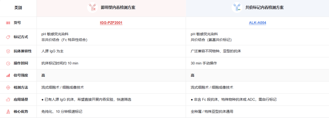
工具一 |pHintra™ 抗体内吞检测试剂(Cat. No.IGG-PZF2001)
pH敏感的荧光染料已标记Fab片段,能特异性识别抗体的 Fc 段、不干扰抗原结合,10min即可形成稳定的抗体复合物,适用于流式细胞术或细胞成像技术等多种方法。
工具二 |pHintra™ 抗体标记试剂盒(Cat. No.ALK-A004)
将pH敏感的荧光染料通过氨基共价标记到自己的抗体上。原理上不依赖抗体的种属与亚型,灵活性更高,手动操作仅需约 30 分钟,同样适用于流式细胞术或细胞成像技术等多种方法。
办案第一步,先确认眼前是否为真案,再锁定作案时间。
用Cat. No. ALK-A004标记 HER2 靶向抗体后,在 HER2 阳性的 SK-BR-3 细胞上,内吞信号增强,呈现时间依赖性(Fig1A);而在两个对照里——靶点阴性的 MDA-MB-468 细胞、以及 IgG1 isotype 对照——均未检测到内吞信号(Fig1B)。说明该实验中的信号来自靶点介导的内吞,而非非特异性吸附。
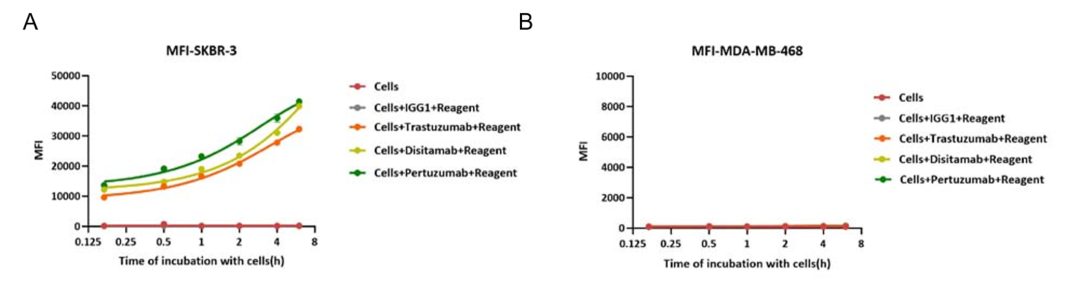
Fig1 Application of pH-Sensitive Fluorescent Dye Antibody Labeling Kit in Endocytosis Detection. The HER2-targeting antibody and its IgG1 isotype control were labeled using the pH-Sensitive Fluorescent Dye Antibody Labeling Kit (Cat. No. ALK-A004) and evaluated for internalization in SK-BR-3 and MDA-MB-468 cells. Robust receptor-mediated internalization of the HER2 antibody was observed in SK-BR-3 cells, with internalization increasing in a time-dependent manner during co-incubation, whereas no internalization was detected in MDA-MB-468 cells or for the IgG1 isotype control.
另一件工具(Cat. No. IGG-PZF2001),也留下了同样的记录(Fig2A、B)。
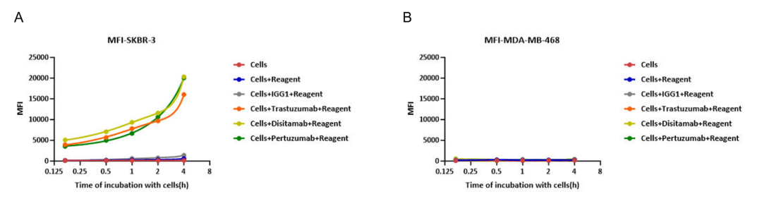
Fig2 In the assessment of ADC efficacy using the Antibody Internalization Detection Reagent (Cat. No. IGG-PZF2001) with HER2-positive SK-BR-3 cells and HER2-negative MDA-MB-468 as target cells; (A) Prolonged co-incubation of HER2-targeting antibodies with HER2-positive SK-BR-3 cells enhanced antibody internalization. (B) whereas no internalization was observed in HER2-negative MDA-MB-468 cells.
抗体偶联上毒素、换装成 ADC,它进入细胞的表现还和原来一样吗?这是 ADC 研发者最常追问的问题之一。两件工具给出了一致的参考:
-
Cat. No. ALK-A004:在不低于 0.01 μg/mL 时,trastuzumab-based 与 disitamab-based ADC 的内吞信号,与对应裸抗相当(Fig3A、B);
-
Cat. No. IGG-PZF2001:在不低于 0.1 μg/mL 时,两款 ADC 均检出剂量依赖的内吞信号,其中 disitamab 组 ADC 与裸抗未观察到统计学显著差异(Fig4A、B)。
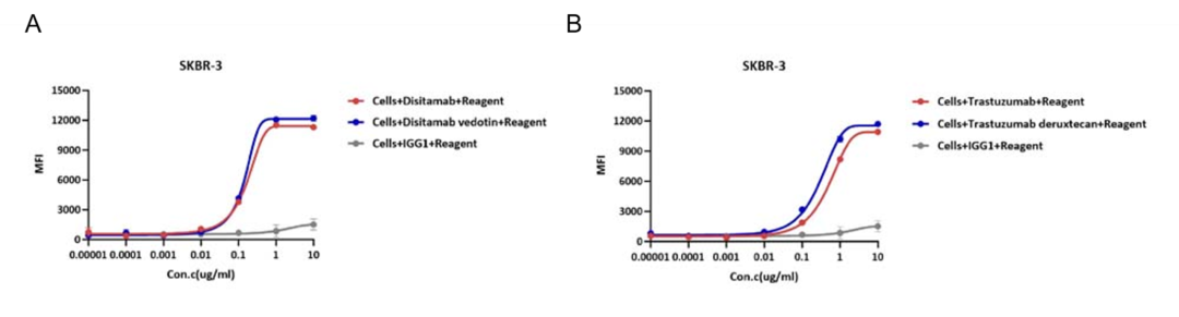
Fig3 Application of pH-Sensitive Fluorescent Dye Antibody Labeling Kit in Endocytosis Detection. (A-B) The HER2-targeting antibodies (trastuzumab and disitamab), their corresponding ADCs, and an IgG1 isotype control were labeled with a pH-sensitive fluorescent dye using the Antibody Labeling Kit (Cat. No. ALK-A004), and their cellular internalization was assessed in SK-BR-3 cells. Both HER2-targeting antibodies exhibited efficient internalization at concentrations ≥ 0.01 μg/mL in a dose-dependent manner, whereas no detectable internalization was observed below this threshold. Furthermore, both the trastuzumab-based and disitamab-based ADCs showed levels of internalization comparable to those of their respective naked antibody counterparts.
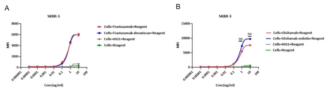
Fig4 In the assessment of ADC efficacy using the Antibody Internalization Detection Reagent (Cat. No. IGG-PZF2001) with HER2-positive SK-BR-3 cells as target cells; (A) The trastuzumab-based ADC effectively internalizes relative to its naked antibody counterpart at concentrations ≥0.1 μg/ml, demonstrating dose-dependent enhancement with comparable efficacy between the two. (B) Disitamab-based ADC effectively internalizes relative to its naked antibody counterpart at concentrations ≥0.1 μg/ml, demonstrating dose-dependent enhancement, with no statistically significant difference observed between the two.
Cat. No. ALK-A004采用氨基共价标记技术,原理上不依赖对抗体种属的识别。
人源、小鼠重组、兔重组的 Anti-CD20 抗体,标记后在 CD20 阳性的 Raji 细胞上均检出内吞信号(Fig5A、B、C)。这支持对不同来源抗体进行直接标记与检测。
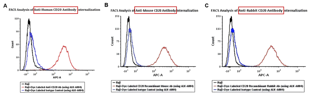
Fig5 FACS Analysis of Anti-CD20 Antibody Internalization. (A) Anti-Human CD20 Ab, (B) Anti-CD20 Recombinant Mouse Ab, and (C) Anti-CD20 Recombinant Rabbit Ab, together with their corresponding isotype controls, were labeled using pH-Sensitive Fluorescent Dye Antibody Labeling Kit (Cat. No. ALK-A004). Raji cells were treated with the dye-labeled antibodies and isotype controls separately at 37℃ for 4 hours, then analyzed by flow cytometry. APC signal was used to evaluate the internalization signal.
除了经典靶点,这套方法对新型抗体结构是否同样适用?
Cat. No. IGG-PZF2001验证了双特异性抗体 trontinemab——它带有一个转铁蛋白受体(TfR)穿梭模块,可借 TfR 介导的转运穿过血脑屏障,目前处于阿尔茨海默病临床开发阶段。在 HEK293/人 TfR 稳定细胞系上共孵育 4 小时,检出内吞信号(Fig6)。说明此方法对新型抗体结构同样适用。
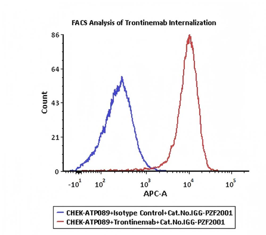
Fig6 Trontinemab and Human IgG1 isotype control were labeled with Antibody Internalization Detection Reagent (Cat. No. IGG-PZF2001). HEK293/Human Transferrin R Stable Cell Line were treated with Trontinemab-Internalization Detection Reagent conjugate and Isotype control-Internalization Detection Reagent conjugate separately for 4 hours, then analyzed by Flow cytometric. APC signal was used to evaluate the activity (Routinely tested).
案件的最后一环,是找到抗体的落脚点。
SK-BR-3细胞经溶酶体绿色荧光探针、pHintra 红色标记抗体与核染料三重染色后成像,数据显示:抗体相关的红色信号与溶酶体标记存在共定位(Fig7A–D)。这为抗体进入溶酶体相关区室,提供了空间成像证据。
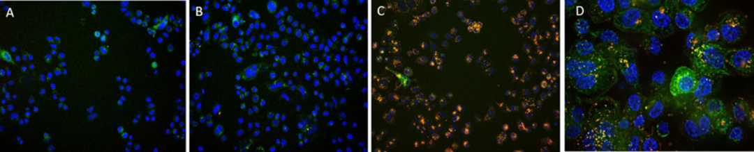
Fig7 SK-BR-3 cells were treated with CellLights Lysosome GFP (green) for 16 hours followed by treatment with Anti-Her2 Abs-Internalization Detection Reagent conjugate and IgG1 Isotype-Internalization Detection Reagent conjugate separately for 16 hours (red), then stained with NucBlue Live ReadyProbes (blue) for 20 minutes and imaged on the EVOS M7000. A. Antibody Internalization Detection Reagent (Cat.No.IGG-PZF2001). B. IgG1 Isotype-Internalization Detection Reagent conjugate. C. Anti-Her2 Abs-Internalization Detection Reagent conjugate. D. Anti-Her2 Abs-Internalization DetectionReagent conjugate (Z-stacking).
结案:证据链闭环
从确认信号真伪与时间,到核验换装、跨来源协查、接手新型案件,再到最终的空间定位——五件证据环环相扣,构成一条完整的内吞证据链:信号真伪与时间(特异性 + 时间动力学)→ 剂型对比(ADC vs 裸抗)→ 不同来源抗体 → 新型抗体结构 → 空间成像。
以上结论均限定于所示的特定分子与实验条件。它们回答的是这套方法可以如何验证内吞,而您自己的抗体表现如何,还需亲手验证。
选型速查

ACROBiosystems
inquiry@acrobiosystems.com
15117918562
(备注:姓名+公司)